Accurately diagnosing a condition or injury is critical to creating a successful treatment plan.
Comprehensive testing helps to diagnose an injury or condition which enables our physicians to create the most effective treatment plan. The benefits of these tests, highlighted below, are that all of them are done on-site in our fully-equipped, state-of-the-art facilities, making it both efficient and convenient for you.
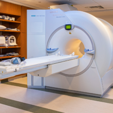
MRI
Magnetic resonance imaging (MRI) is a diagnostic procedure that uses a combination of a large magnet, radiofrequencies, and a computer to produce detailed images of organs and structures within the body. The MRI creates a magnetic field around you, then pulses radio waves to the area of your body to be pictured. The radio waves cause your tissues to resonate. A computer records the rate at which your body’s various parts (tendons, ligaments, nerves) give off these vibrations, and translates the data into a detailed, two-dimensional picture. MRI, a non-invasive way to look inside of the body, does not use ionizing radiation, unlike X-rays or computed tomography (CT scans). An MRI is helpful at diagnosing many common orthopedic problems. For example, the sensitivity of MRI in diagnosing an ACL tear is about 90%. MRI is the chosen method for imaging the soft tissues of the musculoskeletal system, such as ligaments, tendons and cartilage. These structures are most commonly injured during athletic and traumatic events. MRI can be also be used for imaging the spine, hip, shoulder, elbow, wrist, knee, ankle, and foot.
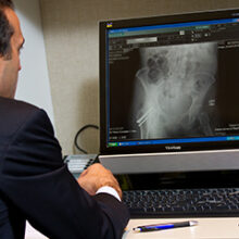
Digital X-ray
A proper x-ray is one of the most crucial pieces of diagnostic information that aides in your diagnosis and treatment. Digital X-ray imaging includes sensors which are used instead of traditional photographic film. This allows immediate image viewing by avoiding chemical film processing and the ability to digitally transfer and enhance images. Digital X-rays also utilizes less radiation to produce an image of similar contrast to conventional x-rays and provides a wider and more dynamic image. Digital x-ray technology allows for special techniques that enhance the overall display of the image. The higher level of accuracy of digital X-ray images also results in a more accurate diagnosis.
Nerve Conduction Study
Injuries or diseases that affect nerves and muscles can slow or halt the movement of the electrical signals transmitted by these nerves and muscles. A nerve conduction study (NCS) measures the speed and degree of electrical activity in your nerves. This allows your doctor to make a proper diagnosis from any symptoms of pain, weakness or numbness you may be experiencing. A nerve conduction study can determine if a nerve is functioning normally. For this test, wires (electrodes) are taped to the skin in various places along the nerve pathway. Then the nerve is stimulated with an electric current. As the current travels down the nerve pathway, the electrodes placed along the way capture the signal and time how fast that signal is traveling. A slower or weaker than optimal signal at any of the points of measurement determines damage to that location in the nerve. This test is commonly used for conditions such as carpel tunnel syndrome and ulnar nerve entrapment. Nerve conduction studies may also be used during treatment to test the progress being made.
DXA Scan
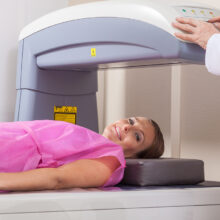
The only test available to diagnose osteoporosis is the bone mineral density (BMD) test, which is also referred to as the DXA scan (which stands for Dual-energy X-ray Absorptiometry). The DXA machine, which employs an extremely small amount of radiation, takes a picture of the bones in the spine, hip, total body and wrist and calculates their density. The test is painless and noninvasive, requiring no special preparations. The value scores reported compare the bone density tested to that of an average young adult. The test results are reported in a number known as a T-score. Diagnosis and treatment plans for bone conditions are dependent on standardized T-scores values. The World Health Organization has established T-score classifications as normal, osteopenia (reduced bone mass, less severe than osteoporosis), and osteoporosis (literally meaning “porous bones”, fragile bones). Treatment recommendations depend on into which of these categories your T-score falls.
UOA’s Diagnostic Testing Department is uniquely distinguished in the area of skeletal health. Patricia Seuffert, Research Coordinator and Nurse Practitioner at UOA, won the 2012 Professional Award presented by NJ’s Health and Senior Services and NJ Interagency Council for Osteoporosis for her work on improving hip fracture management with hospitalized patients. In addition, UOA is one of only two ISCD-accredited facilities in the state of New Jersey.
