Evaluating Shin Pain

Appropriate evaluation and diagnostic imaging is important to determine the cause and extent of injury for individuals experiencing chronic shin pain. Appropriate clinical assessment begins with a thorough patient history that includes an assessment of:
- Pain
- Symptoms
- Function
- Training history
- Prior history of bone stress injury
The clinician should conduct a thorough clinical examination which often includes tests to identify injury. As part of our desire to understand bone stress injuries, UOA has formally studied which tests are most sensitive and specific to identify bone stress injury. Palpation of the shin to identify the location and extent of pain is important. Individuals who experience pain, decreased height and loss of balance while doing a single leg hop test have been identified as having a high likelihood of bone stress injury.
Imaging and Shin Pain
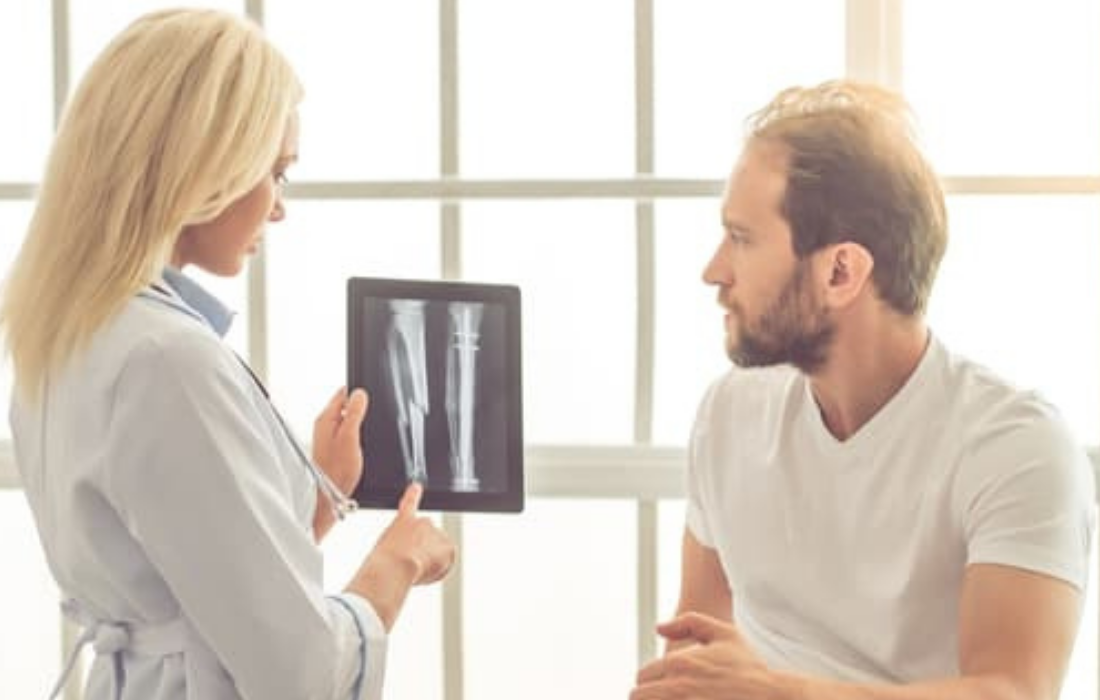
Use of radiography (X-ray) has been noted to be an important first step for patients who experience shin pain by the American College of Radiologists Appropriateness Criteria (ACRAC) for use of imaging. Though findings in the initial X-ray evaluation may be low (3 to 30 percent), there is value in doing an initial X-ray to rule out other causes of pain such as tumor, infection or fracture.
A follow-up X-ray two to three weeks later is also recommended by the ACRAC, as evidence of bone healing may be visualized at this point. It is important to note that less than 50 percent of serial (repeat) X-rays will ever be positive with individuals who suffer a bone stress injury and this can create a false negative finding.
UOA recently completed a study to evaluate the value of an X-ray during the initial clinical exam and compared X-ray findings with MRI scans. Results of our study showed that those with evidence of low level bony stress injury on MRI did not show evidence of injury on an initial X-ray, while higher levels of bony stress injury showed increasing evidence on X-ray. Our study has been submitted for formal publication in a peer-reviewed journal and it further substantiates the value of an initial X-ray for those individuals who experience more than one week of shin pain.
Individuals who have a negative X-ray but a strong clinical finding may be referred for additional imaging such as a bone scan or MRI. Bone scan involves a higher dose of radiation and is often contraindicated for most adolescents. MRI is the gold standard for imaging individuals with a negative X-ray but a high suspicion of bone stress injury.
Appropriate treatment can begin when a true diagnosis is established and a course of treatment and rehabilitation is laid out by your physician. If you have shin pain, request an appointment at UOA to get on the road to recovery.

